Beyond thick glasses,
your retinal health is our concern.
Lifelong management beyond simple vision correction
High Myopia Total Care Solution
Ultra-high myopia patients
ultimately choose Doctor ICL's 'specialness' for a reason
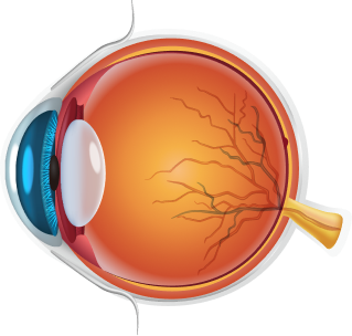
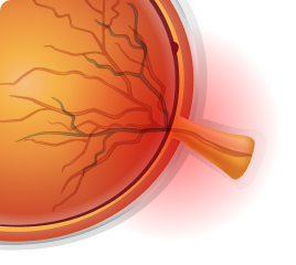
Normal Eye (Normal spherical eyeball)

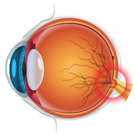
(Ultra-)High Myopia (Elongated eyeball)
High myopia is not simply a state of poor vision.
The eye becomes structurally weakened,
a condition requiring lifelong careful monitoring.
That is why Doctor ICL manages not only before and after lens implantation, but also lifelong potential complications.
Elongated eyeball, thinned tissues...
Causes of complications.
Glaucoma
Increased risk of optic nerve damage as the eyeball elongates
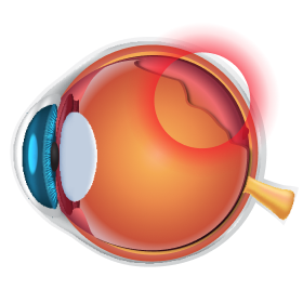
Retina
Risk of holes or detachment as the retina thins

Cataract
Earlier cataracts than average due to structural weakening
Doctor ICL understands the patient's eyes best.
We manage not only vision correction but also lifelong potential
complications.
From lens implantation to cataracts, glaucoma, and retinal diseases.
We will be your
'lifelong trusted eye doctor'.
Stretched optic nerve requires
more detailed monitoring.
Glaucoma in high myopia patients
is much more difficult to identify than usual.
Doctor ICL clearly identifies these subtle differences.
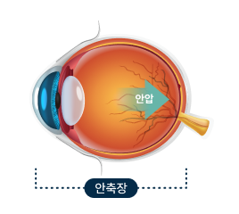
Eye Elongation
Axial length increases
Optic Nerve Deformation
Optic nerve gets stretched
Optic Nerve Vulnerability
Damage risk even at normal eye pressure
High myopia often causes the optic nerve shape itself to be
deformed,
making it difficult to detect glaucomatous damage
with standard examinations.
Is it a change from myopia,
or damage from glaucoma?
Doctor ICL clearly distinguishes through
precise analysis and cross-verification.
Structural Analysis
Detecting subtle thickness changes
Precisely analyzes the thickness of the optic disc, retinal nerve, and fiber layer.
Functional Verification
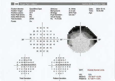
Confirming actual visual field damage
Double-checks structural abnormalities and functional damage through visual field testing.
From identifying vulnerabilities before surgery to preemptive defense after surgery
Before lens implantation, we identify optic nerve vulnerabilities in advance,
and continue regular eye pressure checks and optic nerve monitoring after surgery.
Beyond thick glasses,
your retinal health is our concern.
Lifelong management beyond simple vision correction
High Myopia Total Care Solution
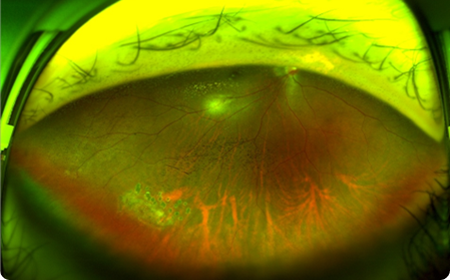
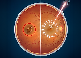
Thorough coverage of invisible areas —
Ultra-Dense Barrier Laser
Retinal detachment prevention — the difference is in 'density'.
Standard Laser
Doctor ICL Laser
Doctor ICL's 'Ultra-Dense Barrier Laser'
forms a seamless defense even in invisible areas.
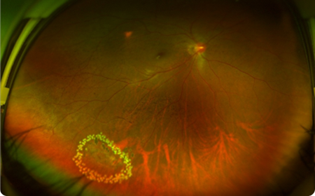
Finding the
'hidden 20%' that standard exams cannot reach.
Standard Fundus Imaging
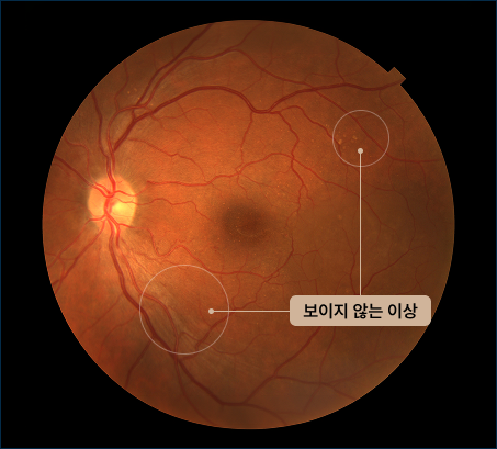
Cannot detect peripheral diseases
Deep-Scan
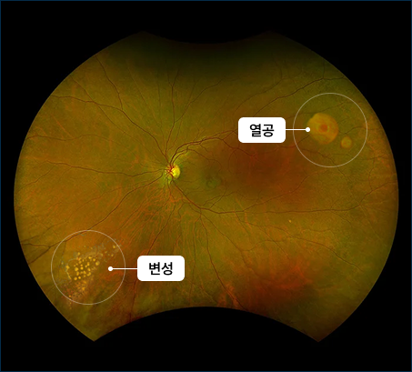
Detects peripheral tears/degeneration
For high myopia with high retinal detachment risk, problems always hide in the 'periphery'.
We scan even invisible blind spots with university hospital-grade wide-angle equipment.
Doctor ICL
Ultra-Dense Laser
3 Safety Standards
01Zero-Gap Targeting
Captures weakened areas such as retinal tears and lattice degeneration
with 3-4 layers of surrounding coverage.
02High-Density Barrier
Applies laser at much higher density than standard dosage
to physically block disease spread.
03Long-term Retical Guard
Before lens implantation, firmly secures vulnerable retinal areas
to preemptively block potential retinal disease at the same site
and create a safe surgical environment.
“Cataract surgery —
vision recovery is a given.”
For high myopia, 'lifelong safety' must also be planned.
Doctor ICL considers the safety of the retina and optic nerve for decades after surgery.
For high myopia with elongated eyeballs,
standard calculation methods are insufficient.
The eyeball of high myopia,
abnormally elongated to focus.
This structural change dramatically increases
the difficulty of measuring power during cataract surgery.

Elongated Eyeball
Requires specialized design optimized for the structure

Differentiated Surgery
A completely different approach from standard cataract surgery

Error-Free Results
Overcome with Doctor ICL's exclusive power calculation method
The risk of post-surgical vision error
caused by elongated axial length —
Resolved with Doctor ICL's precise data technology.
Accuracy (Calculation)
POINT 01. Precise Power Calculation
“The longer the eye, the greater the potential error.”Standard formulas cannot accommodate axial length variables.
We apply high myopia-specific IOL Calculation to bring target vision error close to zero.
Lens Selection (Selection)

POINT 02. Custom Lens Matching
“The optimal selection guide for sensitive eyes”High myopia eyes are sensitive to light scatter due to thin retinas.
We recommend premium lenses that minimize
astigmatism correction and nighttime glare,
tailored to individual characteristics.
Safety (Safety)
POINT 03. Low-Stimulation Technique
“Advanced low-stimulation protocol to compensate for insufficient tissue durability”High myopia eyes have weak structures supporting the lens, making them susceptible to damage from even small pressure changes during surgery.
Doctor ICL minimizes the burden on the capsular bag and supporting tissues with precise low-stimulation surgical techniques, reducing the risk of unexpected damage and secondary complications.
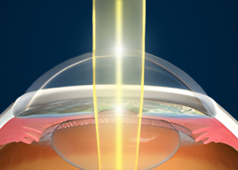
For high myopia, rather than
'reshaping' the cornea, we must
'preserve' it.
Standard LASIK/LASEK (Corneal Ablation)
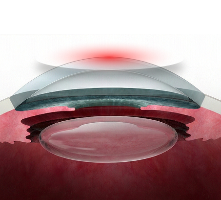
Structural Deformation
Doctor ICL (Corneal Preservation)
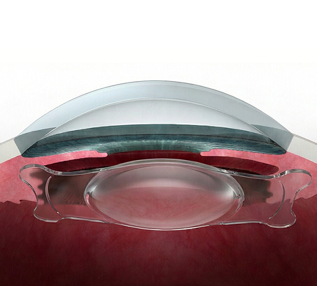
Structural Preservation
The first condition for safe vision correction is 'preservation'.
Doctor ICL does not damage your cornea by even one micron.
We don't operate unconditionally.
Structural stability is confirmed first,
then surgery proceeds.
Space Analysis
Foundation Reinforcement
Precision Insertion
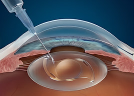
We thoroughly analyze the space for the lens,
reinforce the weakened retina first, then proceed with surgery.
Doctor ICL insists only on 'world-class standards'.

[ 3 KEY FEATURES ]
“No Iridotomy Required”
Facilitates aqueous flow to prevent glaucoma complications
“Biocompatible Material”
Special material the body doesn't reject — zero inflammation/foreign body sensation
“Simultaneous Astigmatism Correction”
Clearly resolves even high astigmatism with a single lens
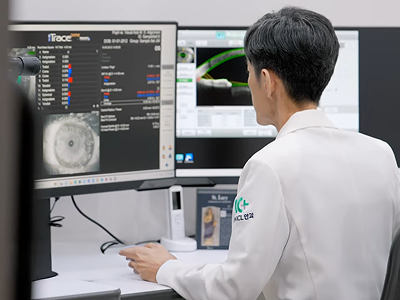
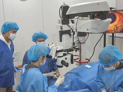
Surgery ends in 15 minutes, but data is recorded for a lifetime.
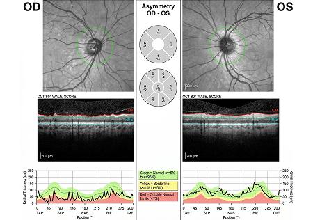
Spectralis OCT
High-resolution anterior segment CT scannerSpectralis OCT precisely images the retina and choroid layer by layer to identify structural changes.
Repeated imaging of the same area enables accurate comparison with previous exams, with even subtle changes tracked in accumulated data.
This examination is used for early detection and long-term follow-up of optic nerve and retinal diseases including glaucoma, macular degeneration, and retinal disorders.
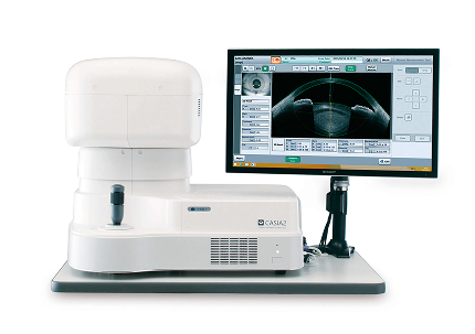
TOMEY CASIA2
High-resolution anterior segment CT scannerTOMEY CASIA2 captures the anterior segment with high-resolution OCT to precisely examine the cornea, lens, and anterior chamber structure.
Records the anterior segment structure in a non-contact manner during examination, creating objective data for structural changes before and after lens implantation.
The acquired anterior segment data is used for pre-surgical baseline setting as well as post-surgical structural stability comparison and follow-up.
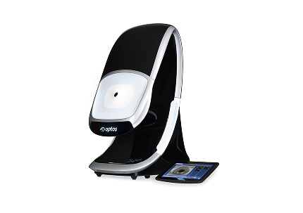
OPTOS-Daytona
Non-mydriatic ultra-widefield fundus cameraCaptures 200 degrees (approximately 80%) of the entire retina without dilation, enabling rapid examination of central and peripheral retina.
Enables early diagnosis of previously undetected peripheral retinal diseases (retinal tears, degeneration, etc.).
As a non-mydriatic device, there is no waiting time for dilation, and normal activities are possible immediately after examination.
Spectralis OCT
High-resolution anterior segment CT scannerSpectralis OCT precisely images the retina and choroid layer by layer to identify structural changes.
Repeated imaging of the same area enables accurate comparison with previous exams, with even subtle changes tracked in accumulated data.
This examination is used for early detection and long-term follow-up of optic nerve and retinal diseases including glaucoma, macular degeneration, and retinal disorders.
TOMEY CASIA2
High-resolution anterior segment CT scannerTOMEY CASIA2 captures the anterior segment with high-resolution OCT to precisely examine the cornea, lens, and anterior chamber structure.
Records the anterior segment structure in a non-contact manner during examination, creating objective data for structural changes before and after lens implantation.
The acquired anterior segment data is used for pre-surgical baseline setting as well as post-surgical structural stability comparison and follow-up.
From pre-surgery examination to lifelong management,
5 STEP System
- Step 1. [Deep-Scan] Ultra-precise retina/optic nerve examination (OCT/Optos)
- Step 2. [Preemptive Defense] Priority laser treatment upon retinal tear/degeneration discovery
- Step 3. [Custom Surgery] Lens power calculation & insertion matched to elongated eyeball
- Step 4. [Double Check] Final confirmation of lens position and eye pressure (glaucoma)
- Step 5. [Lifelong Management] Data-based follow-up of retina, optic nerve, and anterior segment structure